Pictures from Hysteroscopy Surgery of Normal and Abnormal Uterine Cavities
Investigating Problems Related to Infertility and/or Miscarriage
Hydrosalpinx and IVF success rates
- Hysteroscopy is a procedure done by a gynecologist or infertility specialist physician that investigates uterine causes of infertility and miscarriages.
- It is done by passing a narrow scope through the vagina and cervical opening to visualize the inside of the uterine cavity.
- There are various abnormalities that can be found that can interfere with initial embryo implantation, or with ongoing pregnancy.
- These structural abnormalities of the uterine cavity can prevent pregnancy from beginning or they can prevent continuation of pregnancy (increasing the risk for miscarriage).
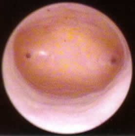
Hysteroscopy picture of a normal uterine cavity
Tubal ostia (openings) seen on each side
<>
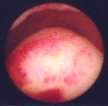
Office hysteroscopy (2.7 mm diameter scope) was done to view the uterine cavity
Substantial endometrial polyp bulging up into the cavity at lower part of image
<>
Same uterine cavity as above - after the polyp was resected with operative hysteroscopy
<>
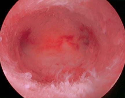
Pillars of uterine scar tissue are at the top of the uterine cavity in this photo
This woman had a D&C for heavy bleeding following delivery of a baby
<>
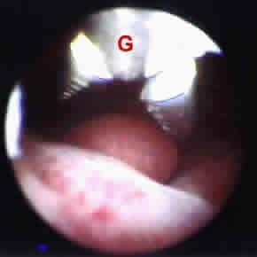
Small polyp being resected with office hysteroscopy, grasper at "G"
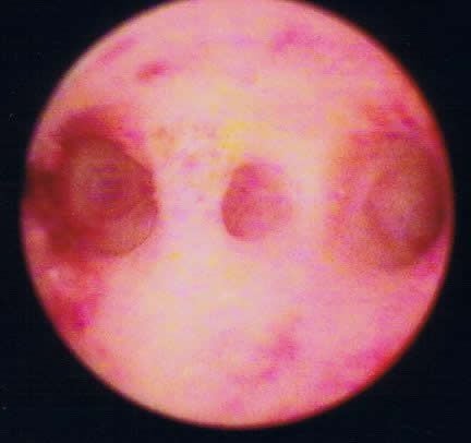
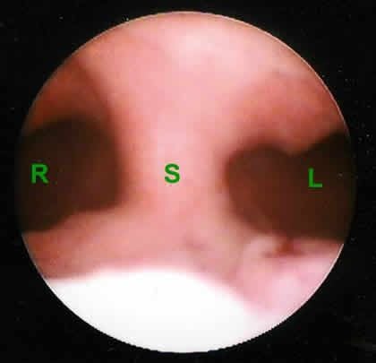
Uterine septum at "S" in middle of picture
Right side of cavity at R and left side at L
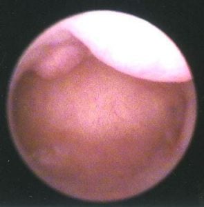
2 fibroid tumors are shown bulging down into the endometrial cavity
These fibroids were resected with surgery - resulting in a normal uterine cavity
Categories
About the AFCC Blog
Welcome to the Advanced Fertility Center of Chicago’s blog! Here, you will find information on the latest advancements in fertility care and treatments, including IVF, IUI, third-party reproduction, LGBTQ+ family building, preimplantation genetic testing, and more. Since 1997, we’ve used our experience and continuous investment in the latest fertility technology to help thousands of patients grow their families. Contact us today for more information or to schedule a new patient appointment.


