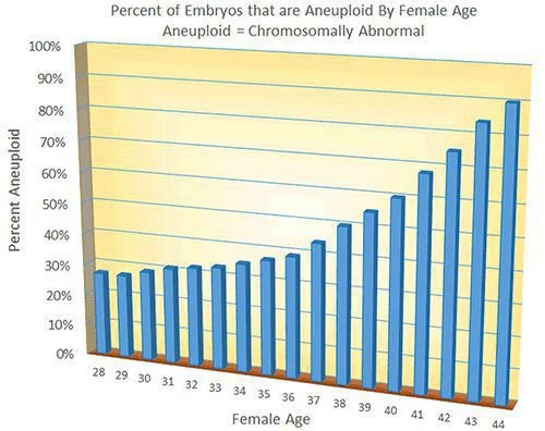
The main cause of increased risk for miscarriage in “older” women is increased rates of chromosomal abnormalities in their eggs.
Background
There are 2 general types of chromosomal abnormalities:
- Numerical abnormalities where there is an abnormal number of chromosomes is called aneuploidy.
- Such as a missing (monosomy) or an extra chromosome (trisomy). An example of monosomy is Turner Syndrome. A well known example of trisomy is Down Syndrome.
- Structural abnormalities where there is a problem with the structure of a chromosome.
- Examples include translocations, duplications and deletions of part of a chromosome
Aneuploid eggs and embryos are also responsible for most of the decline in fertility with female aging and for the low pregnancy success rates with IVF for women over 40.
The increased rate of chromosomal abnormalities in women of advanced reproductive age has been well documented in research studies.
- Preimplantation genetic screening, PGS, can be done to test embryos for chromosomal abnormalities before they are transferred back to the uterus
- Using PGS we can almost completely eliminate chromosomally abnormal embryos from the pool of embryos being considered for transfer
- If we have a normal embryo for transfer this significantly increases the chance for a successful pregnancy
- See our IVF pregnancy success rates
Diagnosis of endometriosis
The only way to be sure whether a woman has endometriosis is to perform a surgical procedure called laparoscopy that allows us look inside the abdominal cavity with a narrow scope.
Sometimes we strongly suspect that the disease is present based on the woman’s history of very painful menstrual cycles, painful intercourse, etc., or based on the physical examination of the woman or ultrasound findings.
Rate of chromosomally abnormal human eggs and embryos by age
Chromosomal Problems in Aging Eggs
We do not know exactly why there is an increase in chromosomal abnormalities in the eggs of women as they age. However, research studies have clarified some of the issues involved.
The meiotic spindle is a critical component of eggs that is involved in organizing the chromosome pairs so that a proper division of the pairs can occur as the egg is developing. An abnormal spindle can predispose to development of chromosomally abnormal eggs.
Study on Human Egg Spindles and Female Age
An excellent study published in the medical journal “Human Reproduction” in October of 1996 investigated the influence of maternal age on meiotic spindle assembly in human eggs.
- This study gives insight into how chromosomally abnormal eggs (and therefore, embryos) are more common in older women.
- 17% of the eggs studied from women 20-25 years old were found to have an abnormal spindle appearance and at least one chromosome displaced from proper alignment.
- 79% of the eggs studied from women 40-45 years old were found to have an abnormal spindle appearance and at least one chromosome displaced from proper alignment.
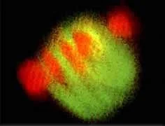
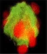
The pictures below are from this journal article. These photos were taken with confocal fluorescence microscopy of eggs stained with special dyes to show the spindles and chromosomes.
- The orange spots are pairs of chromosomes in the eggs.
- The green area is the spindle apparatus.
Chromosomally Normal Egg
Egg from a woman in her 20’s
Chromosomes line up straight on spindle
Chromosomally Abnormal Egg
Egg from a woman in her early 40’sChromosomes line up erratically – a mishap during chromosome separation is much more likely
When the chromosomes line up properly in a straight line on the spindle apparatus in the egg, the division process would be expected to proceed normally so that the egg would end up with its proper complement of 23 chromosomes.
However, with a disordered arrangement on an abnormal spindle, the division process may be uneven – resulting in an unbalanced chromosomal situation in the egg. Older eggs are significantly more likely to have abnormally functioning spindles – which causes an increased rate of chromosomal problems in the mature eggs.
Why do some women have more chromosomally abnormal eggs than average for their age?
- Possible insight from a study about a gene that regulates normal egg development (in mice)
In August 2009 a study was published (article by Lelanda referenced below) showing that mice that had a mutation in a specific gene had significantly higher rate of chromosomal abnormalities than normal.
- The gene is called Bub1
- The gene is involved in regulation of the proper division of pairs of chromosomes during the egg maturation process
- The degree of the chromosomal problems were found to increase with maternal age
- The chromosomal abnormalities were passed on to the embryos and resulted in high rates of loss after implantation
- It is not yet known how often a gene mutation like this might cause similar problems with human reproduction
- You might think we are very different from mice – but we are not (sorry about that)
This study is very interesting and could give insight into reasons for some women having infertility, recurrent IVF failure, or recurrent miscarriages.
- Similar research needs to be done in humans
- In the future we could possibly have a blood test (genetic testing) that could predict risks for chromosomal problems in a woman’s eggs and embryos.
Chromosomal Errors in Eggs Causing Down Syndrome – Screening in Pregnancy
- Screening options in early pregnancy for Down Syndrome have changed over the last several years
- Pregnant women that are age 35 and older are now commonly offered testing with cell free fetal DNA by their OB/GYN doctor
- This involves a blood test on the woman at about 10 weeks of pregnancy
- DNA fragments called “cell-free DNA” in the woman’s blood are analyzed
- Most cell-free DNA is from the mother but some of it comes from the placentaThe cell-free
- DNA is analyzed to see if there is an increased risk for the baby having Down Syndrome (trisomy 21) or trisomy 13 or 18
- This technology detects about 98-99% of cases of Down syndrome with a false-positive rate under 0.5%
- However, It is less accurate for detecting trisomy 13 and 18
Sources:
- D.E. Battaglia, et al: Influence of maternal age on meiotic spindle assembly in oocytes from naturally cycling women. Human Reproduction October, 1996; (Vol. 11): Pages 2217-2222.
- S. Lelanda, et al: Heterozygosity for a Bub1 mutation causes female-specific germ cell aneuploidy in mice. PNAS August, 2009; Pages 12776-12781.
- The American College of Obstetricians and Gynecologists (ACOG) Practice Bulletin #77, “Screening for Fetal Chromosomal Abnormalities.


