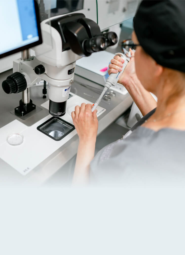
PGD, Preimplantation Genetic Diagnosis for Genetic Disorders

Preimplantation Genetic Testing for Monogenic Defects (PGT-M)

Genetic Testing for Single Gene Defects

Pre-implantation genetic diagnosis is a technique that is used along with in vitro fertilization, IVF and allows testing of embryos for certain characteristics such as their chromosomal makeup and also testing for genetic diseases that are passed on through families. When one or both partners in a couple are carriers of the genetic mutation that could leave could lead to a serious medical condition in the child in vitro fertilization and preimplantation genetic diagnosis testing can be performed on their embryos. In past years couples that carried a genetic mutation could choose between not having children or having prenatal testing done with either amniocentesis at about 16 to 18 weeks of pregnancy or with chorionic villus sampling at about 11 to 12 weeks to see if the baby was affected with the genetic condition. The couple then has the option of terminating the pregnancy if the baby is affected with the disorder. Now couples that are carriers for genetic diseases have the option of having IVF and PGD to screen their embryos prior to transferring them to the uterus. Not all diseases can be tested for in this manner. Single gene disorders are caused through the inheritance of a defective gene. These disorders are classified as either recessive or dominant. A recessive disorder requires 2 bad copies of the gene to pass the disease on to the baby. With a dominant single gene disorder only one copy of the defective gene is needed to cause the disease. There are over 1000 single gene disorders that have been identified at the present time. Many of these disorders are very rare. However, some are so common in certain ethnic groups that routine screening to check whether someone is a carrier is recommended prior to getting pregnant. This is often referred to as carrier genetic testing (or screening).

The most common single gene disorders that PGD has been used for are:

  • Cystic fibrosis
  • Tay-Sachs disease
  • Spinal muscular atrophy (SMA)
  • Hemophilia
  • Sickle cell disease
  • Duchennes muscular dystrophy
  • Thalassemia
However, there are hundreds more genetic diseases that can have single gene testing done using IVF and PGD. A partial list of relatively common single gene diseases is below.

Autosomal recessive disorders

Sanhoff disease, Gaucher disease, adenosine Deaminase deficiency, glycogen storage disease, Fanconi anemia, adrenal hyperplasia, phenylketonuria (PKU).

Autosomal dominant disorders

Neurofibromatosis, Von-Hippel Lindau, myotonis dystrophy, Huntington’s Disease, Marfan syndrome, osteogenesis imperfecta, Charcot-Marie-Tooth, APP early onset Alzheimers, polycystic kidney disease, retinitis pigmentosa, familial adenomatous polyposis, achondroplasia.

X-linked disorders

Ornithine carbamyl transferase deficiency, Fragile X, X-linked hydrocephalus.

How is IVF and PGD with genetic testing done?

In vitro fertilization is performed, the embryos have trophectoderm biopsies done on day 5 and 6 at the blastocyst stage of development. The biopsied cells are then sent to the genetics laboratory for evaluation. The genetics laboratory does their fancy genetics magic and determines whether the embryo contains the “abnormal” DNA of the genetic disease in question, or not. The testing is done utilizing high tech genetics technology that analyzes the DNA sequence in the cells for the specific gene in question.

PGD genetic testing for cystic fibrosis

An example of using PGD for genetic testing for single gene defects is the situation where a couple are both carriers for the recessive gene that causes cystic fibrosis. In order for a child to have cystic fibrosis it needs to get one copy of the abnormal recessive gene from each parent. A normal gene combined with an abnormal gene does not cause the disease. That person would only be a carrier and would not affected by cystic fibrosis. If both parents are carriers of this recessive genetic condition then every child they have has a one in 4 risk for having the cystic fibrosis disease. Therefore, sometimes a couple that are both known carriers for the abnormal cystic fibrosis gene will decide to undergo in vitro fertilization with PGD genetic testing of their embryos for cystic fibrosis.
    • In such a case we expect that one out of 4 embryos would have both of genes being abnormal which would lead to a child with the cystic fibrosis disease
    • Those abnormal embryos would not be transferred back to the mother’s uterus
  • We expect 2 out of 4 embryos to be carriers for the abnormal gene (one normal copy and one abnormal)
  • We expect 1 out of 4 to have 2 copies of the normal gene (not carriers)
  • Embryos would be chosen for transfer to the uterus that genetic testing showed did not have 2 copies of the abnormal cystic fibrosis gene.

Blastomere biopsy on day 3 after fertilization is one way that PGD testing can be done

With blastomere biopsy, a slit is made in the shell of the embryo on day 3 and a pipette is introduced into the embryo. Then one of the cells is carefully teased out of the embryo which would have approximately 8 cells at that time. If it’s an 8 cell embryo and we remove one cell for testing, it then is a 7 cell embryo. At this stage of development, all of the cells are undifferentiated. Therefore, losing one cell would not take anything away from the subsequent child. The cells are all clones of each other at this point and would continue dividing and eventually differentiate into specific cell types. The biggest problem with day 3 blastomere biopsy is that it has been shown to result in a significantly reduced implantation potential after the embryo is transferred to the uterus.

Trophectoderm biopsy on day 5 or 6 at the expanded blastocyst stage is a better way to do embryo biopsy

This is a newer technique that utilizes biopsy at a later stage of embryonic development. At the blastocyst stage there has been some differentiation of the cells into an inner cell mass (destined to become the fetus) and the trophectoderm (precursor to the placenta). A small portion of the trophectoderm cells are removed for the biopsy. The inner cell mass is not disturbed. A disadvantage of trophectoderm biopsy is that with current technology it takes about 24 hours to get results back from the biopsy. Therefore, since the embryos should be transferred by day 5 or early on day 6 it is not currently practical to do trophectoderm biopsy and transfer fresh embryos to the uterus. Therefore, trophectoderm biopsy is performed and the embryos are frozen and subsequently thawed and transferred later after results of testing are available. Trophectoderm biopsy is less traumatic to the embryos than blastomere biopsy and therefore results in a higher success rate.

Choose AFCC for Preimplantation Genetic Diagnosis in Chicago

Is PGD right for you? Our team is here to answer all your questions and concerns so that you can move forward with confidence. Contact Advanced Fertility Center of Chicago today for more information about PGD or to schedule a consultation at one of our fertility clinics in Chicago, Downers Grove, or Gurnee, IL.

Questions?

We are here to answer any questions or concerns you may have so that you feel completely confident when taking the first step toward building your family.

Share on facebook
Share on twitter
Share on linkedin

The site uses cookies, pixels and other similar technologies, as further described in our privacy statement. By using our site, you agree to our use of cookies.