Intrauterine Adhesions, Asherman's Syndrome - Scar Tissue In Uterine Cavity
What is Asherman's syndrome?
The condition of scarring within the endometrial cavity of the uterus is often referred to as Asherman's Syndrome.
A normal uterine cavity and endometrial lining is necessary in order to conceive and maintain a pregnancy. Scar tissue within the uterine cavity can partially or completely obliterate the normal cavity and can interfere with conception, or increase the risk for miscarriage or other complications later in the pregnancy.
Causes of Asherman's Syndrome
- It is most commonly caused by the trauma to the lining from a D&C (dilation and curettage)
- A recently pregnant uterus is much more susceptible to developing Asherman's after D&C compared to a non-pregnant uterus
- It is sometimes caused by scarring after uterine surgeries such as Cesarean section or myomectomy for fibroid tumors
- In rare cases it is caused by infection, such as genital tuberculosis (rare in the US)
- We have seen some women with uterine adhesions that have never had any D&C or other surgery - some cases of Asherman's are of unknown cause
Asherman's is uncommon
- It has been estimated that about 1% of D&Cs will cause intrauterine scarring
- Having multiple D&Cs increases the risk for developing scar tissue in the uterus
- D&C done for retained placental tissue postpartum is much more likely to cause Asherman's.
Some studies estimate the risk after postpartum curettage to be as high as 25%
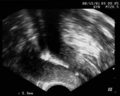
Ultrasound picture showing bright (hyperechoic) uterine lining - scar tissue in uterine cavity
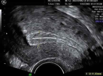
For comparison, an ultrasound picture of a uterus with a normal endometrial lining
The endometrium here is 11.2 mm thick (yellow cursors)
A normal endometrial thickness is about 8 to 15 mm
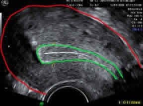
Same image showing the outer uterine contour outlined in red and the "triple stripe" of 3 layers in the uterine endometrial lining outlined in green. The cervical canal is well visualized at lower right.
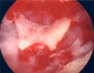
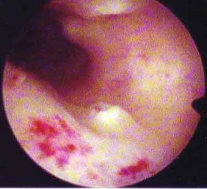
View of the inside of a scarred uterine cavity using hysteroscopy
Thick white scar tissue is seen (same woman as in ultrasound at top of this page)
White patches are the scarred areas of adhesions, pink is normal tissue, dark red is blood
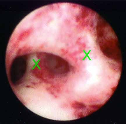
Office hysteroscopy showing 2 bands of scar tissue (at green X's) going from "floor" to "ceiling" in uterine cavity
Same view after in-office hysteroscopic resection of the scar tissue
<strong>This uterus appeared normal when studied with ultrasound alone</strong>
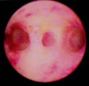
Another photo of scarring of endometrium
Area of left tubal opening at 3 o'clock
Area of right tubal opening at 9 o'clock
Scarred region is in the middle
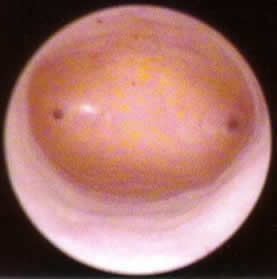
Normal hysteroscopy view for comparison
Intrauterine adhesions can be cut during hysteroscopy to improve chances for embryo implantation (pregnancy) and to reduce the risk of miscarriage.
Categories
About the AFCC Blog
Welcome to the Advanced Fertility Center of Chicago’s blog! Here, you will find information on the latest advancements in fertility care and treatments, including IVF, IUI, third-party reproduction, LGBTQ+ family building, preimplantation genetic testing, and more. Since 1997, we’ve used our experience and continuous investment in the latest fertility technology to help thousands of patients grow their families. Contact us today for more information or to schedule a new patient appointment.


